Along with the rapid development of modern technology, advanced dental techniques have gradually been born to meet the needs of both patients and doctors during examination and treatment at dental facilities. Dental X-ray is considered one of the indispensable techniques for the dental industry today. This method helps the treatment to take place more quickly, accurately and effectively when it can produce images of pathological signs that cannot be seen with the naked eye.
1. What is a dental X-ray?
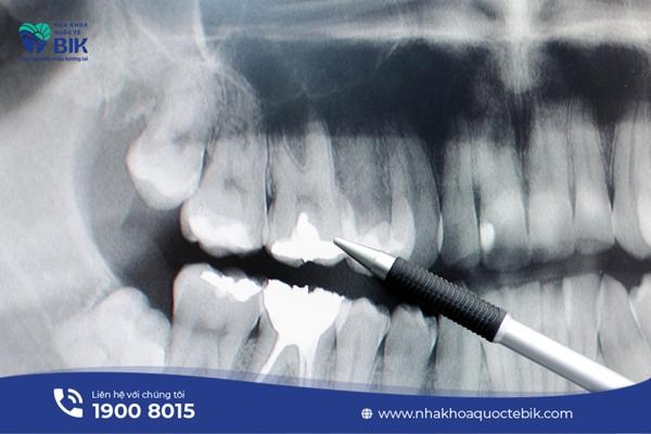
A dental X-ray is a technique that uses X-rays to record images of the oral cavity accurately and realistically. This image will show all parts including teeth, jawbones, soft tissues, tooth roots, dental pulp, etc. on a film or a digital sensor. Thanks to these images, doctors can easily check and detect dental problems from deep inside such as tooth decay, tooth abscess or even tumors or other abnormal conditions.
2. What are the effects of dental X-rays?
Dental X-rays are gradually becoming an indispensable technique for doctors when treating oral diseases because this technique brings benefits to both doctors and patients:
– Doctors have a general image to limit possible complications when performing porcelain crowns, braces, dentures, etc.
– Observing even the details deep under the tooth root such as the nerve canal helps the tooth extraction surgery to take place in the safest way.
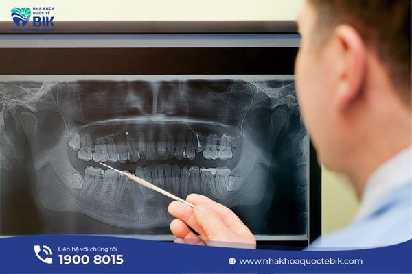
– Because they have the most general and detailed image of the oral cavity, doctors will soon detect lesions in the jawbone area that are difficult to see. Not only that, because it can determine the severity of diseases such as periodontitis, tooth decay, pulpitis, gingivitis, etc., doctors can make timely recommendations and treatments for patients.
– Based on the X-ray film, the doctor will determine the position and direction of tooth growth, thereby providing a treatment regimen to improve the aesthetics of the customer.
3. When is it necessary to take a dental X-ray?
Cases that require the use of dental X-ray techniques include:
– Need to check for problems such as: tooth decay, broken teeth, damage to the jaw bone, pulpitis, check jaw bone density…
– Teeth growing crooked or growing through the gums
– The patient feels prolonged toothache
– Before braces
– Before root canal surgery, molar extraction, dental implants, dentures,..
– Observe the development of children’s teeth
4. What are the dental X-ray techniques?
Depending on the need to diagnose oral health conditions, there will be many different X-ray techniques:
4.1. X-ray of a single tooth
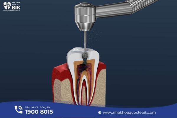
X-ray of a single tooth is a fairly common technique in which the amount of X-rays is limited to the maximum but still ensures enough dose to detect damaged teeth as well as adjacent teeth and still ensures the quality of the resulting film image. The technique of taking X-rays of a single tooth is often applied in cases of tooth decay, need for tooth filling, root canal treatment, etc.
4.2. 3D Dental X-rays
Along with the remarkable development of modern technology, dental clinics also use 3D dental X-rays – a combination of rotating X-ray equipment and modern digital equipment. This technique helps doctors observe structures that cannot be seen with the naked eye. When performing this method, the X-ray image will be a 3D image, showing soft tissues, muscles, jaw bones, nerve tubes, blood vessels, etc.
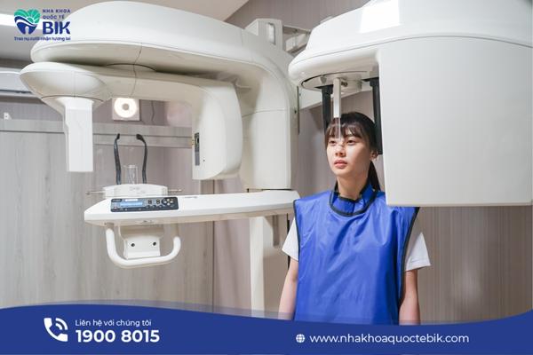
4.3. Panoramic dental X-ray
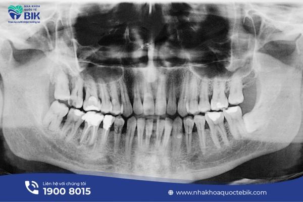
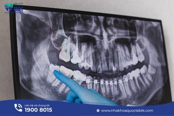
Panorama X-ray helps to record sharp images of the entire oral cavity with high resolution, this image will provide an overview of the jaw, teeth, gums, nasal sinuses and temporomandibular joints. In particular, the panoramic dental X-ray method helps doctors observe the entire two dental arches on the same film, thereby diagnosing diseases such as crooked teeth, impacted teeth, cysts, dental abscesses, etc.
When performing a panoramic dental X-ray, the patient will be wearing a protective cover and asked to bite on a fixed plastic plate on the machine. When the machine starts working, the X-ray bulb and the sensor plate (film) will move in opposite directions, rotating around the patient’s lower jawbone, the patient will also be asked to stand still for about 12-15 seconds while the machine works.
The only drawback of the panoramic dental X-ray technique is that it cannot detect cavities or other problems such as fractures, infections, etc.
4.4. Periapical X-rays
Periapical X-rays will be prescribed in cases where it is necessary to find dental problems under the gums or in the jaw that are difficult to see with the naked eye such as cysts, molars, boils, etc. This technique will take 14-21 films to provide the most accurate images of the entire tooth, from the incisors to the tooth roots, the bone supporting the tooth, etc.
4.5. Perioral X-ray
When taking a perioral X-ray, the patient does not need to put the film in the mouth but only needs to sit up straight in the chair, the X-ray machine will automatically rotate around the patient to record general images of the jawbone and teeth.
This technique is very useful in detecting abnormalities that cannot be observed in both upper and lower jaws, thereby easily detecting oral diseases that are difficult to detect with the naked eye.
4.6. Peri-bite X-ray
With peri-bite X-ray technique, the doctor can see how the teeth touch each other, whether the teeth of both jaws are aligned or not. In addition, the film image produced by this technique also shows the condition of bone loss due to infection or severe gingivitis…
4.7. Bite X-ray
Bite X-ray technique is often indicated when it is necessary to search for foreign objects in the oral cavity because it will produce images of the floor of the mouth or palate. The film will detect pimples, abnormal tissue growth, cleft palate, etc.
5. What should I do before taking a dental X-ray?
A dental X-ray will be prescribed by the doctor depending on the oral condition, so not everyone has to do it. No matter what dental X-ray technique is prescribed, there is no need to prepare anything special before taking the photo.
In the case of tooth extraction, you should eat and drink enough before taking the photo and then clean your mouth thoroughly because after tooth extraction, you will only be allowed to eat soft foods, which can make the patient tired.
You should bring the old X-ray film and medical records (if any) so that the doctor can easily compare and give appropriate advice.
6. Dental X-ray process in dentistry
Dental X-ray is divided into 2 main stages:
6.1. During X-ray
When entering the X-ray room, the doctor will instruct the patient to hold a piece of cardboard or plastic in their mouth to keep the X-ray film steady while taking the photo. At this time, the X-ray machine will surround the head to record images inside the oral cavity.
During the photo, the patient must follow the instructions given, sit still during the photo to avoid overlapping images, blurring, and having to do it again.
6.2. After taking the X-ray
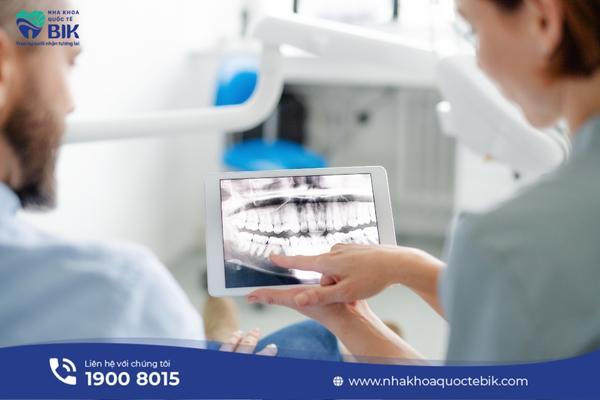
The dental X-ray process is very quick and does not cause any discomfort to the patient. After taking the dental X-ray, the patient only needs to wait for the results and listen to the doctor’s advice. In addition, patients do not need to abstain from any food after taking the film.
7. Subjects who should not have dental X-rays
Although dental X-rays today have been improved and upgraded a lot compared to before. Although the radiation dose of modern digital dental X-ray machines is very low, it is still limited to the following cases of patients:
7.1. Children should not have dental X-rays
When it is necessary to check the tooth growth process or to detect cavities, children may also be prescribed X-rays. In this case, lead protective devices will be equipped to absorb scattered X-rays, minimizing negative impacts on children’s health.
7.2. Pregnant women should not have dental X-rays

In the case of pregnant women, the necessity of having an X-ray will be considered, because although the dose of X-rays is very low, it still has a negative impact on the health of the mother and baby. In case it is necessary to take an X-ray, safety measures for the mother and fetus will be prepared such as a lead shirt or a lead bib to cover the abdomen.
With the information provided above, BIK International Dental Clinic has provided you with all the information you need to know about dental X-ray techniques at the dentist. This seems to be an indispensable technique and a powerful assistant to support doctors throughout the examination, consultation and treatment process.